Adult liver tumor
The incidence of adult liver tumor has been increasing in recent years, which is a severe threat to human health. Liver neoplasm is divided in to benign neoplasm (tumor-like lesions) and malignant neoplasm. The latter include primary liver neoplasm and metastatic liver neoplasm. The anatomy of the liver could be destroyed by the swelling/infiltrative growth of neoplasm or intrahepatic metastasis. The morphology of tumor liver is diverse among different diseases, various stages of the same disease and interindividual variability.
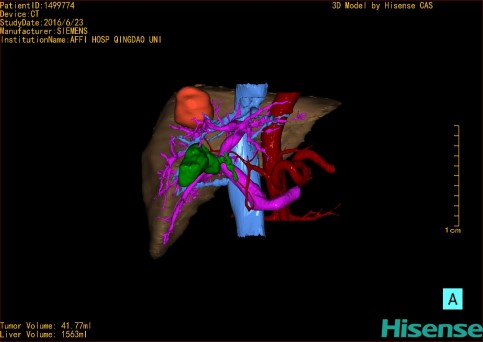
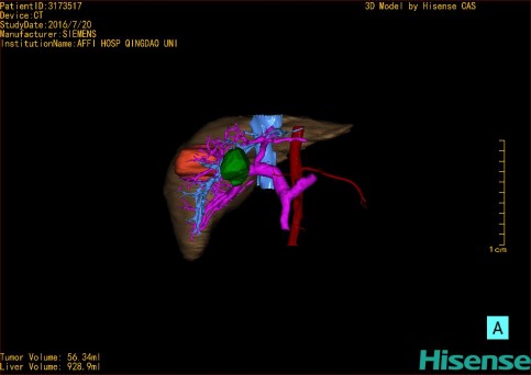
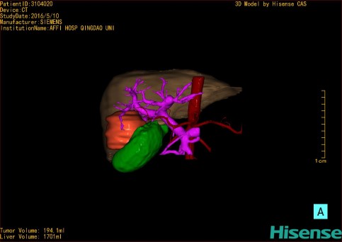
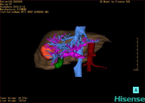
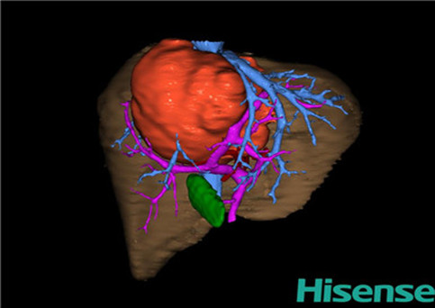
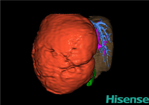
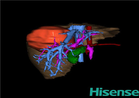
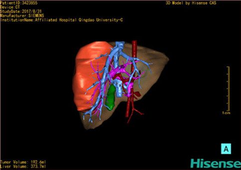
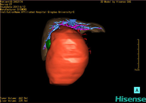
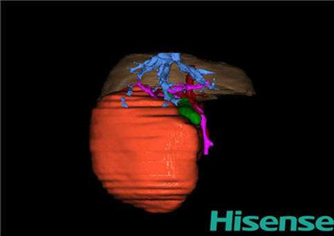
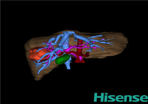
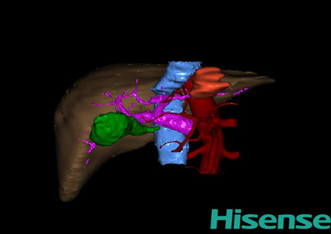
By using Computer Assisted Surgery System (CAS) to load liver tumor CT data set in format of DICOM, a transparent, visible and arbitrary three-dimensional digital model including adult liver parenchyma, neoplasm, portal venous system and hepatic arterial system, hepatic vein and biliary system could be constructed. Thus the digital liver of an individualized adult liver tumor is formed. The digital liver is capable of presenting the functional liver volume and morphology of adult patients, the morphology and distribution of tumors, and the degree of damage to hepatic vascular system and biliary system, etc., which provides individualized clinical information for liver function reserve evaluation, preoperative evaluation, treatment options selection (surgery, radiotherapy, chemotherapy, etc.), evaluation for liver transplantation and prognosis and is beneficial to the individualized and precise treatment.