With the development of medical imaging, 3D reconstruction technology, the innovation of liver surgery and the proposal of precision hepatectomy, the intra-hepatic liver vascular anatomy and the liver segmentectomy based on vascular anatomy was well developed. With the big data analysis of 3D digital liver, we proposed a new way of liver classification system: Dong's digital liver classification system. Professor Dong Qian, from the Affiliated hospital of Qingdao University, analyzed anatomy of thousands of human digital livers from newborn to aged human beings, trying to build a new system of liver classification based on intrahepatic vascular anatomy.
【Introduction】
1260 cases of normal human liver were reconstructed into 3-D digital liver with their DICOM file. According to the sectorial branch divides into different portal branches suppling segments, we built our Dong's digital liver classification system.
Dong's Digital Liver Segments Classification
Type A (8 Segments Type): According to the direction and branches of the intrahepatic portal vein, this type is divided into eight segments, accounting for about 42.62%.
Type B (9 Segments Type): According to the direction and branches of the intrahepatic portal vein, this type is divided into nine segments, accounting for about 36.83%.
Type C (RP Type): According to the direction and branches of the intrahepatic portal vein, this type is divided into seven segments(C-a)or eight segments(C-b), accounting for about 8.09%.
Type D (special variant Type): This type contains several special variant branches of the intrahepatic portal vein, accounting for about 12.46%.
【Principle of Dong's Digital Liver Classification System】
According to the sectorial branch divides into different portal branches suppling segments, we built our Dong's digital liver classification system. Here is some basics component of our digital liver classification system.
The nomenclature of portal vein branches
1st grade portal vein: The main portal trunk, indicating the portal vein from the confluence of superior mesenteries vein and splenic vein to where the portal dividing into right main branch and left main branch.
2nd grade portal vein: From the end-point of 1st grade portal vein to the point where it divides into branches, indicating the right portal vein and left portal vein originate from the main portal trunk.
3rd grade portal vein: The part of portal vein which separate small branches perfusing different liver segment, including umbilical portion of portal vein in the left liver, and right anterior sectorial branch and right posterior sectorial branch in the right liver.
4th grade portal vein: The small branches which perfuse liver segment, which is the basic anatomical resection unit for precise liver resection and also the unit for our Dong's Digital Liver Segments Classification.
The 4th grade portal vein will separate thinner 5th or 6th grade branches. They usually perfuse only small area which is not so useful for hepatectomy.
Video 1 3D simulation showing the portal vein
Principle of Dong's digital liver segment classification
1. Dong's digital liver segment is based on the sectorial branch divides into different portal branches suppling segments, give consideration to the derange of hepatic vein.
2. The 4th grade portal vein is the basic unit of Dong's digital liver segment classification.
3. The portal vein perfusing caudate lobe is especially different from 4th grade portal vein. There are 3-6 small branches originate from the back of right and left portal vein, deranged by 5-8 tiny short hepatic vein. During operation, it is difficult to control the perfusing blood of caudate lobe by Pringle manoeuvre. So, we prefer to define the region as one segment, segment I.
4. To respect the tradition, we defined the area mostly composed of the caudate lobe as segment I. Start from segment II of left upper area of the left liver, we defined the liver as segment II-IX in the clockwise direction.
5. As the variation of left portal vein are relatively few, the segment II, III and IV were defined same with Couinaud. However, the portal veins of right liver vary a lot, which can not be included by only one (segment) classification method.
【Dong's Liver Segments】
According to our big data analysis, we found that few variation occurred in the portal vein of left liver, perfusing segment II, III and IV. However, there are so many variations that we can not sum up into one classification of segmentation. According to the sectorial branch divides into different portal branches suppling segments, we built our Dong's digital liver classification system. We settled the variation into four main types: Type A. 42.62%; Type B: 36.83%; Type C (8.09%): C-a, 4.52%; C-b, 3.57%; Type D, 12.46%
Type A: 8 Segments, accounting for about 42.62%.
Segment I (3 to 6 P1 branches): Caudate lobe. There are 3-6 small branches (P1) originate from the back of right and left portal vein, deranged by 5-8 tiny short hepatic vein.
Segment II, III: Left portal vein divides into third grade portal vein (P2 and P3) respectively perfusion upper and lower outsides of left liver, which is segment II and III.
Segment IV: Portal veins divided from left portal vein perfuse the inner part of left liver, which is segment IV.
Segment V: The right portal vein divides into the right anterior and posterior branches, and then the anterior trunk will further divide into several branches. The branches that perfuse the area of inner lower part of the right liver, which is segment V.
Segment VI: The right posterior portal vein will further separate into right anterior (P6) and posterior branches (P7). The anterior branches perfuse segment VI, lower outside of the right liver.
Segment VII: The right posterior portal vein will further separate into right anterior (P6) and posterior branches (P7). The posterior branches perfuse segment VII, upper outside of the right liver.
Segment VIII (P8): The right portal vein divides into the right anterior and posterior branches, and then the anterior trunk will further divide into several branches. The branches that perfuse the area of upper and inner-superior part of the right liver (P8), which is segment VIII.
Dong's Liver Segments,Type A, 8 Segments Type

Figure 1:Dong's Liver Segments Type A Figure 2:Different view of Type A
Video 2 Dong's Liver Segments Type A
Segment I (3 to 6 P1 branches): Caudate lobe. There are 3-6 small branches (P1) originate from the back of right and left portal vein, deranged by 5-8 tiny short hepatic vein. Volume of Segment I takes about 7% of whole liver volume.

Figure 3:Portal vein branch for segmentⅠof Type A Figure 4:SegmentⅠof Type A
Segment II: Left portal vein divides into third grade portal vein (P2 and P3) respectively perfusion upper and lower outsides of left liver, which is segment II and III. Segment III takes about10% of whole liver volume.

Figure 5:Portal vein branch for segmentⅡof Type A Figure 6:SegmentⅡof Type A
Segment III: Left portal vein divides into third grade portal vein (P2 and P3) respectively perfusion upper and lower outsides of left liver, which is segment II and II. Segment III takes about 8% of whole liver volume.
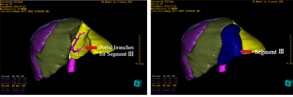
Figure 7:Portal vein branch for segment Ⅲ of Type A Figure 8:Segment Ⅲ of Type A
Segment IV: Portal veins divided from left portal vein perfuse the inner part of left liver, which is segment IV, taking about 16% of whole liver volume.
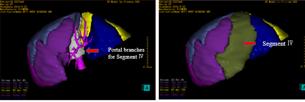
Figure 9:Portal vein branch for segment Ⅳ of Type A Figure 10:Segment Ⅳ of Type A
Segment V: The right portal vein divides into the right anterior and posterior branches, and then the anterior trunk will further divide into several branches. The branches that perfuse the area of inner lower part of the right liver, which is segment V. Segment V takes about 16% of whole liver volume.
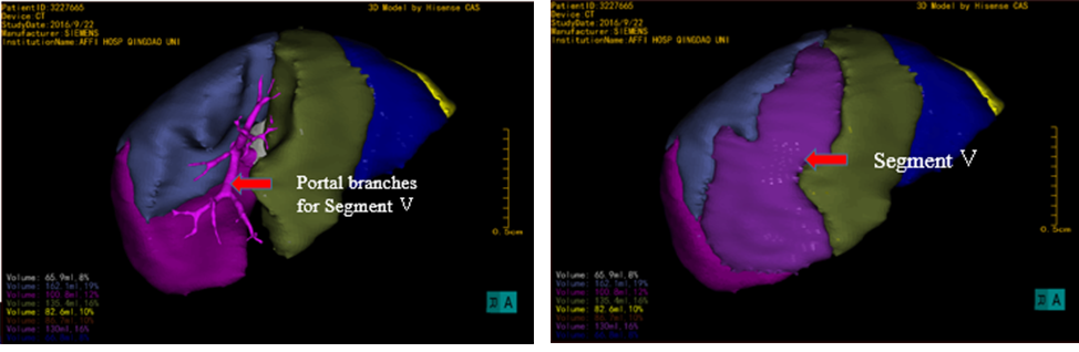
Figure11:Portal vein branch for segment V of Type A Figure 12:Segment V of Type A
Segment VI: The right posterior portal vein will further separate into right anterior (P6) and posterior branches (P7). The anterior branches perfuse segment VI, lower outside of the right liver. Segment VI takes about 12% of whole liver volume.

Figure 13:Portal vein branch for segment Ⅵ of Type A Figure 14:Segment Ⅵ of Type A
Segment VII: The right posterior portal vein will further separate into right anterior (P6) and posterior branches (P7). The posterior branches perfuse segment VII, upper outside of the right liver. Segment VII takes about 10% of whole liver volume.
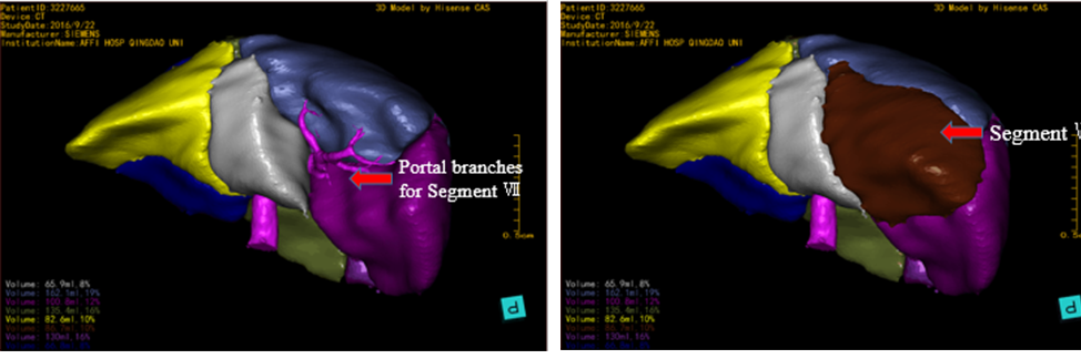
Figure15:Portal vein branch for segment Ⅶ of Type A Figure 16:Segment Ⅶ of Type A
Segment VIII (P8): The right portal vein divides into the right anterior and posterior branches, and then the anterior trunk will further divide into several branches. The branches that perfuse the area of upper and inner-superior part of the right liver (P8), which is segment VIII. Segment VIII takes about 19% of whole liver volume.
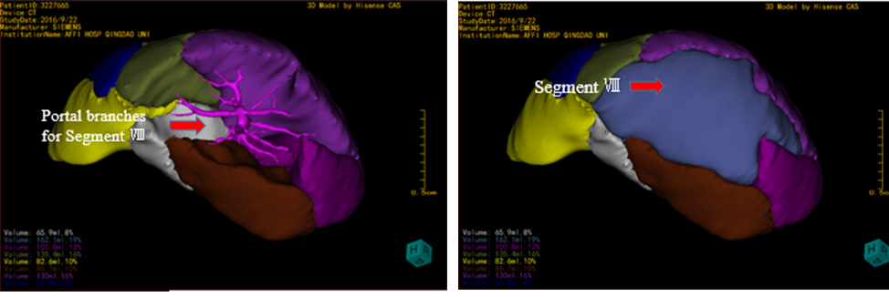
Figure17:Portal vein branch for segment Ⅷ of Type A Figure 18:Segment Ⅷ of Type A
Type B: 9 Segments, accounting for about 36.83%.
Segment I (3 to 6 P1 branches): Caudate lobe. There are 3-6 small branches (P1) originate from the back of right and left portal vein, deranged by 5-8 tiny short hepatic vein.
Segment II, III: Left portal vein divides into third grade portal vein (P2 and P3) respectively perfusion upper and lower outsides of left liver, which is segment II and III.
Segment IV: Portal veins divided from left portal vein perfuse the inner part of left liver, which is segment IV.
Segment V: The right portal vein divides into the right anterior and posterior branches, and then the anterior trunk will further divide into several branches. The branches that perfuse the area of inner lower part of the right liver, which is segment V.
Segment VI: The right posterior portal vein will further separate into right anterior (P6) and posterior branches (P7). The anterior branches perfuse segment VI, lower outside of the right liver.
Segment VII: The right posterior portal vein will further separate into right anterior (P6) and posterior branches (P7). The posterior branches perfuse segment VII, upper outside of the right liver.
Segment VIII (P8): The right portal vein divides into the right anterior and posterior branches, and then the anterior trunk will further divide into several branches. The branches that perfuse the area of upper part of the right liver (P8), which is segment VIII.
Segment IX (P9): The right portal vein divides into the right anterior and posterior branches, and then the anterior trunk will further divide into several branches. The branches that perfuse the area of inner-superior part of the right liver (P9), which is segment IX.

Figure 19:Dong's Liver Segments Type B Figure 20:Different view of TypeB
Video 3 Dong's Liver Segments TypeB
Segment I (3 to 6 P1 branches): Caudate lobe. There are 3-6 small branches (P1) originate from the back of right and left portal vein, deranged by 5-8 tiny short hepatic vein. Volume of Segment I takes about 9% of whole liver volume.

Figure21:Portal vein branch for segment Ⅰ of Type B Figure 22:Segment Ⅰ of Type B
Video 4 Segment Ⅰ of Type B
Segment II: Left portal vein divides into third grade portal vein (P2 and P3) respectively perfusion upper and lower outsides of left liver, which is segment II and III. Segment III takes about 11% of whole liver volume.
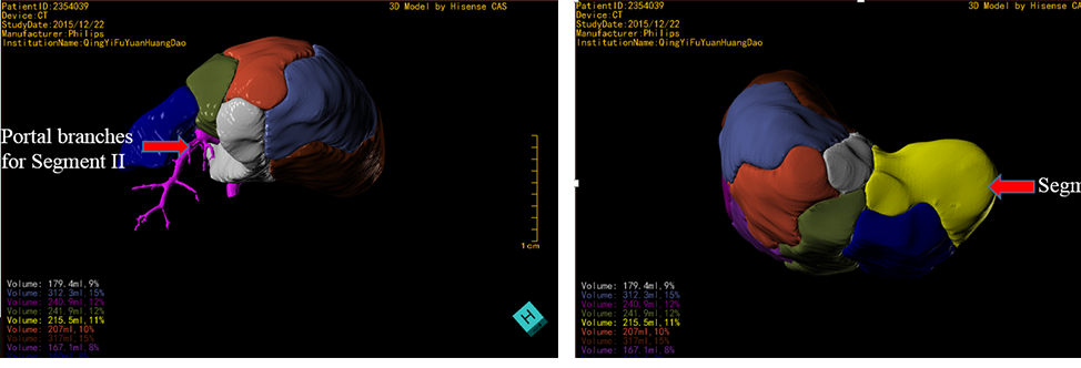
Figure23:Portal vein branch for segment Ⅱ of Type B Figure 24:Segment Ⅱ of Type B
Video 5 Segment Ⅱ of Type B
Segment III: Left portal vein divides into third grade portal vein (P2 and P3) respectively perfusion upper and lower outsides of left liver, which is segment II and II. Segment III takes about 8% of whole liver volume.
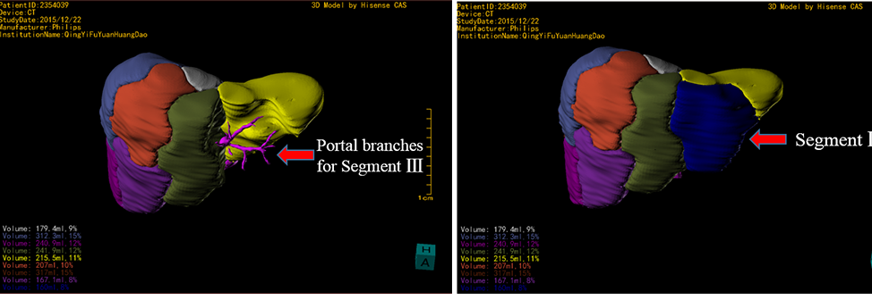
Figure25:Portal vein branch for segment Ⅲ of Type B Figure 26:Segment Ⅲ of Type B
Video 6 Segment Ⅲ of Type B
Segment IV: Portal veins divided from left portal vein perfuse the inner part of left liver, which is segment IV, taking about 12% of whole liver volume.

Figure27:Portal vein branch for segment Ⅳ of Type B Figure 28:Segment Ⅳ of Type B
Video 7 Segment Ⅳ of Type B
Segment V: The right portal vein divides into the right anterior and posterior branches, and then the anterior trunk will further divide into several branches. The branches that perfuse the area of inner lower part of the right liver, which is segment V. Segment V takes about 12.2% of whole liver volume.
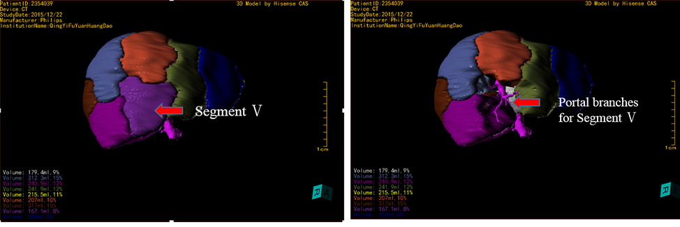
Figure29:Portal vein branch for segment Ⅴ of Type B Figure 30:Segment Ⅴ of Type B
Video 8 Segment Ⅴ of Type B
Segment VI: The right posterior portal vein will further separate into right anterior (P6) and posterior branches (P7). The anterior branches perfuse segment VI, lower outside of the right liver. Segment VI takes about 12% of whole liver volume.

Figure 31:Portal vein branch for segment Ⅵ of Type B Figure 32:Segment Ⅵ of Type B
Video 9 Segment Ⅵ of Type B
Segment VII: The right posterior portal vein will further separate into right anterior (P6) and posterior branches (P7). The posterior branches perfuse segment VII, upper outside of the right liver. Segment VII takes about 15% of whole liver volume.
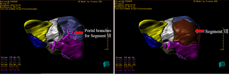
Figure 33:Portal vein branch for segment Ⅶ of Type B Figure 34:Segment Ⅶ of Type B
Video 10 Segment Ⅶ of Type B
Segment VIII (P8): The right portal vein divides into the right anterior and posterior branches, and then the anterior trunk will further divide into several branches. The branches that perfuse the area of upper part of the right liver (P8), which is segment VIII. Segment VIII takes about 15% of whole liver volume.

Figure 35:Portal vein branch for segment Ⅷ of Type B Figure 36:Segment Ⅷ of Type B
Video11 Segment Ⅷ of Type B
Segment IX (P9): The right portal vein divides into the right anterior and posterior branches, and then the anterior trunk will further divide into several branches. The branches that perfuse the area of inner-superior part of the right liver (P9), which is segment IX. Segment VIII takes about 10% of whole liver volume.

Figure 37:Portal vein branch for segment Ⅸ of Type B Figure 38:Segment Ⅸ of Type B
Video12 Segment Ⅸ of Type B
Type C: Right posterior portal vein is arcuate type or not, accounting for about 8.09%.
Right posterior portal vein does not divide into main branches like Type A or B, which separate into 5-11 branches from an arcuate trunk. During precise hepatectomy, it is difficult to resect only the segment VI or VII as type A or type B. The proportion of Type C is not high, but it owns considerable importance in precise surgery. When right posterior portal vein is arcuate type and right anterior portal vein separate only P8 and P5, we define it as Type C-a. When right posterior portal vein is arcuate type and right anterior portal vein separate to P5, P8 and P9, we define it as Type C-b.
Segment I (3 to 6 P1 branches): Caudate lobe. There are 3-6 small branches (P1) originate from the back of right and left portal vein, deranged by 5-8 tiny short hepatic vein.
Segment II, III: Left portal vein divides into third grade portal vein (P2 and P3) respectively perfusion upper and lower outsides of left liver, which is segment II and III.
Segment IV: Portal veins divided from left portal vein perfuse the inner part of left liver, which is segment IV.
Segment V: The right portal vein divides into the right anterior and posterior branches, and then the anterior trunk will further divide into several branches. The branches that perfuse the area of inner lower part of the right liver, which is segment V.
Segment RP (Right Posterior portal vein):Right posterior portal vein does not divide into main branches like Type A or B, which separate into 5-11 branches from an arcuate trunk. During precise hepatectomy, it is difficult to resect only the segment VI or VII as type A or type B. The proportion of Type C is not high, but it owns considerable importance in precise surgery.
Segment VIII (P8): The right portal vein divides into the right anterior and posterior branches, and then the anterior trunk will further divide into several branches. The branches that perfuse the area of upper and inner-superior part of the right liver (P8), which is segment VIII.
When right posterior portal vein is arcuate type and right anterior portal vein separate only P8 and P5, we define it as Type C-a.
Segment IX (P9): The right portal vein divides into the right anterior and posterior branches, and then the anterior trunk will further divide into several branches. The branches that perfuse the area of inner-superior part of the right liver (P9), which is segment IX.
When right posterior portal vein is arcuate type and right anterior portal vein separate to P5, P8 and P9, we define it as Type C-b.
Segment RP (Right Posterior portal vein):Right posterior portal vein does not divide into main branches like Type A or B, which separate into 5-11 branches from an arcuate trunk. During precise hepatectomy, it is difficult to resect only the segment VI or VII as type A or type B. The proportion of Type C is not high, but it owns considerable importance in precise surgery. Segment PR takes about 20% of whole liver volume.

Figure 39:Portal vein branch for segment RP of Type C Figure 38:Segment RP of Type C
Video 13 Dong's Liver Segments Type C
Type D: Special type.
Special type, takes about 12.43%, including variations that cannot classify into any type of the previous three type, including: Right anterior portal vein is divided from left portal vein (51.59%); right anterior portal vein divide into 4-8 small branches perfusing right anterior part of liver (25.97%); P6 is divided from right anterior portal vein; P2 and P3 are from common trunk; right anterior portal vein divided from Sagittal portal vein.
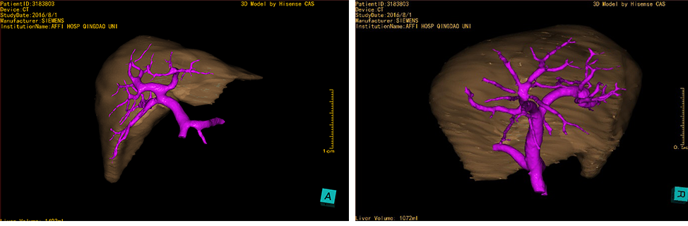
Figure 41:The anterior portal vein in right liver lobe arises from left portal vein stem
Figure 42:The right anterior branches of portal vein simultaneously originates 7 equally thick branches to dominate inner right liver.
Video 14 The anterior portal vein in right liver lobe arises from left portal vein stem
Video 15 The right anterior branches of portal vein simultaneously originates 7 equally thick branches to dominate inner right liver.
Wang Huanmin、Liu Zhimin(Beijing Children's Hospital,Capital Medical University);Li Long、Liu Shuli、Qiao Guoliang、Sun Xuefeng (Capital Institute of Pediatrics);Zheng Shan、Dong Kuiran、Qiao Zhongwei(Children’s Hospital of Fudan University);Lv Yeming、Lv Fan、Liu Ming(Xin Hua Hospital Affiliated to Shanghai Jiao Tong University School of Medicine);Wen Zhe、Liang Jiankun、Ai Bin(Guangzhou Women and Children's Hospital);Liu Juncheng、Yan Chaogui(The First Hospital, Sun Yat-sen University);Zhan Jianghua、Wang Chunxiang(Tianjin Children's hospital);Wei Mingfa、Peng Yang、Zhu Tianqi(TongJi Medical College Hua Zhong University of Science &Technology);Zhang Mingman、Dai Xiaoke、Qin Yong(Children's Hospital of Chongqing Medical College);Shu Qiang、Luo Wenjuan、Liang Jiawei(Children's Hospital of Zhejiang University);Bai Yuzuo、Lu Zaiming(ShengJing Hospital of China Medical University);Li Zhaozhu、Zhao Yanming(The Second Affiliated,Haerbin Medical University);Xi Hongwei、Yang Hui(Children's Hospital of Shanxi Province);Yang Linhong、Chen Changfa、Yao Fengqing、Li Jiade(Linyi Citizen Hospital;Zhu Chengfang、Shan Haibin(Gaomi Citizen Hospital).(names not listed in order)
Thanks to the above cooperation units and professors for the strong support and help of this study!